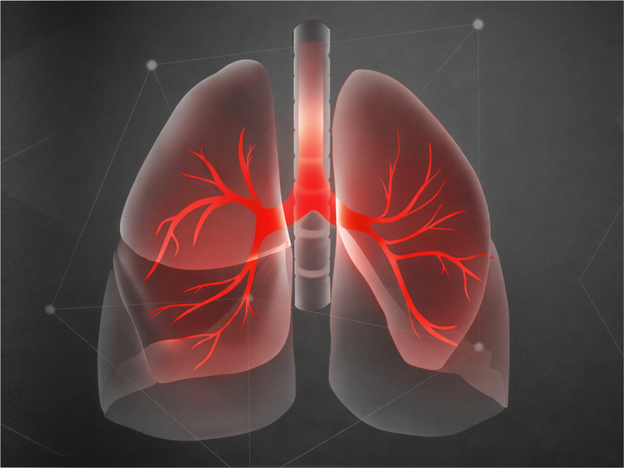
INTERSTITIAL LUNG DISEASE (ILD)
Interstitial lung disease (ILD), also called diffused parenchymal diseases, is a heterogeneous group of disorders characterized by fibrosis (scarring) of the lungs. Hypersensitivity pneumonitis is the most prevalent ILD in India, followed by CTD-ILD and idiopathic pulmonary fibrosis.
The pathogenesis of ILD is complex and is not yet fully understood. It is believed to begin with injury to alveolar epithelial cells often from environmental exposures, smoking or occupational factors in individuals with genetic susceptibility. This injury triggers abnormal wound-healing responses, leading to excessive fibro-proliferation, extracellular matrix (ECM) deposition, immune activation and vascular/endothelial damage.
Myofibroblasts from various sources produce excess ECM proteins like collagen. Injured and overactive alveolar epithelial cells trigger fibroblast activation and chemokine release. These fibroblasts continue to drive fibrosis through cytokines and epigenetic changes. Immune cells such as macrophages and T and B-lymphocytes also become activated and release pro-fibrotic signals. Endothelial cells add to the process by expressing adhesion molecules and chemokines, leading to vascular dysfunction and repair.
The classification system used to describe interstitial lung disease categorizes conditions based on clinical, histopathological or radiologic parameters. Most ILDs share common structural remodeling of the distal airspaces leading to impaired gas exchange.
The common ILDs are as follows:
1. Idiopathic ILDs
The cause of these idiopathic conditions remains elusive, but their progression tends to be characterized by persistent and often worsening fibrosis, functional lung impairment and a declining prognosis.
2. Autoimmune-related ILDs
Chronic disease includes connective tissue disease-associated interstitial lung disease (CTD-ILD). Diseases such as systemic sclerosis and rheumatoid arthritis are CTD-ILD conditions that can cause inflammation and damage to the lungs.
3. Exposure-related ILDs
Some exposure-related diseases are chronic and have a high risk of developing a progressive-fibrosing phenotype, such as hypersensitivity pneumonitis, which is mostly related to the inhalation of organic particles.
4. Other ILDs
Examples include chronic eosinophilic pneumonia, malignant diseases with associated interstitial lung disease (e.g. lymphangitis carcinomatosis) and acute eosinophilic pneumonia. Accurate identification of ILD is critical as it guides targeted treatment, improving patient outcomes and quality of life.
The following is the key laboratory marker used to evaluate and diagnose ILD: